
Randevu +90 324 238 83 83
7/24 Bilgi Hattı. +90 541 720 83 83


Home »
Diabetic retinopathy is a complication of diabetes affecting the eyes. It results from damage to the blood vessels in the light-sensitive tissue at the back of the eye, the retina. Initially, Diabetic Retinopathy may cause no symptoms or only mild visual problems. Eventually, it can lead to blindness.
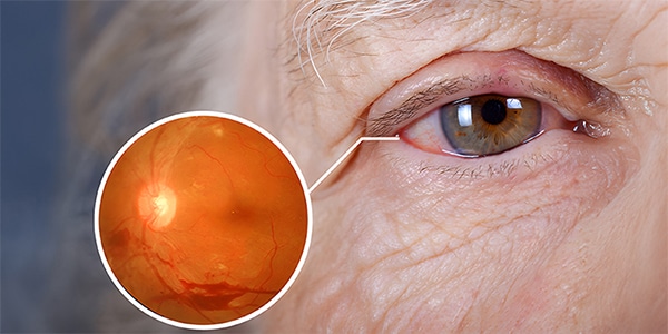
In particular, diabetic retinopathy may have no symptoms in its early stages. As it progresses, you may experience:
Careful diabetes management is the best way to prevent vision loss. If you have diabetes, you need an annual eye examination even if your vision seems fine.
Developing diabetes during pregnancy (gestational diabetes), or having diabetes before pregnancy, increases the risk of diabetic retinopathy. If you are pregnant, your ophthalmologist may recommend additional eye examinations throughout pregnancy.
Contact your ophthalmologist immediately if your vision changes suddenly or becomes blurred, spotty or hazy.
Over time, too much sugar in the blood can block the tiny blood vessels supplying the retina, cutting off blood flow. The eye then tries to form new blood vessels. However, these do not develop properly and can leak easily.
In particular, Early diabetic retinopathy, or non-proliferative diabetic retinopathy (NPDR), is the more common form.
With NPDR, the walls of the retinal blood vessels weaken. Small bulges protrude from the walls of smaller vessels and sometimes leak fluid and blood into the retina. Larger retinal vessels may also begin to dilate and become irregular in diameter. NPDRcan progress from mild to severe as more blood vessels become blocked.
Sometimes, retinal blood vessel damage causes fluid accumulation (oedema) in the central retina, or macula. If macular oedema reduces vision, treatment is needed to prevent permanent vision loss.
Advanced diabetic retinopathy proliferative diabetic retinopathy is the more severe form into which the disease can progress. Damaged blood vessels close off, causing new abnormal vessels to grow in the retina. These vessels are fragile and may leak into the clear, jelly-like substance filling the centre of the eye, the vitreous.
Eventually, scar tissue from new vessel growth can cause the retina to detach from the back of the eye. If the new vessels interfere with normal fluid flow, pressure builds inside the eye. This can damage the optic nerve, which carries images to the brain, causing glaucoma.
Anyone with diabetes can develop diabetic retinopathy. The risk increases with:
Ultimately, Diabetic Retinopathyinvolves abnormal blood vessel growth in the retina. Complications can cause serious visual problems.
Vitreous haemorrhage: new vessels can bleed into the clear, jelly-like substance filling the centre of the eye. If bleeding is slight, you may see only a few dark spots or floaters. In more severe cases, blood may fill the vitreous cavity and completely block your vision.
vitreous haemorrhage alone does not usually cause permanent vision loss. The blood generally clears from the eye within a few weeks or months. Unless the retina is damaged, your vision will probably return to its previous clarity.
Retinal detachment: Abnormal vessels associated with diabetic retinopathy stimulate scar tissue growth, which can pull the retina away from the back of the eye. This can cause floaters, flashes of light or severe vision loss.
Glaucoma: New vessels may form in the front of the eye (iris), obstructing normal fluid flow and causing pressure to build. This pressure can damage the optic nerve, which carries images from the eye to the brain.
Blindness. Diabetic Retinopathy, macular oedema, glaucoma or a combination of these conditions can cause complete vision loss, particularly if poorly managed.
In particular, Diabetic retinopathy cannot always be prevented. However, regular eye examinations, good blood sugar and blood pressure control, and early intervention for visual problems help prevent serious vision loss.
If you have diabetes, your risk of diabetic retinopathy can be reduced by taking the following steps:
Manage your diabetes. Make healthy eating and physical activity part of your daily routine. Aim for at least 150 minutes of moderate aerobic activity, such as walking, each week. Take diabetes medicines or insulin as prescribed.
Monitor your blood sugar. You may need to check and record your blood sugar several times a day, or more frequently if you are ill or stressed. Ask your doctor how often you should test it.
Glycated haemoglobin testing should be discussed with your doctor. The glycated haemoglobin, or haemoglobin A1C, test reflects your average blood sugar over the preceding two to three months. For most people with diabetes, the A1C target should be below 7%.
Keep your blood pressure and cholesterol under control. Healthy eating, regular exercise and losing excess weight help. Medicines may also be needed.
If you smoke or use other tobacco products, stop. Smoking increases the risk of various diabetic complications, including diabetic retinopathy.
Pay close attention to changes in vision. Contact your ophthalmologist immediately if vision changes suddenly or becomes blurred, spotty or hazy.
Remember that diabetes does not necessarily cause vision loss. Taking an active role in managing it helps prevent complications.
In particular, Diabetic Retinopathyis best diagnosed through a comprehensive eye examination. Eye drops dilate the pupils so the doctor can see inside the eyes more clearly.
During the examination, the ophthalmologist checks the internal and external eye structures for abnormalities.
After your pupils are dilated, dye is injected into a vein in your arm. Photographs are then taken as the dye circulates through the blood vessels in your eyes. The images can identify blocked, damaged or leaking vessels.
This test provides cross-sectional images showing retinal thickness. It helps determine how much fluid, if any, has leaked into retinal tissue. Further OCT tests monitor the response to treatment.
Treatment aims to slow or stop progression and depends largely on the type and severity of diabetic retinopathy.
If you have mild or moderate non-proliferative diabetic retinopathy , immediate treatment may not be necessary. Your ophthalmologist will monitor your eyes closely to determine when treatment may be needed.
Work with your diabetes doctor (endocrinologist) to identify ways to improve diabetes management. When Diabetic Retinopathy is mild or moderate, good blood sugar control often slows progression.
proliferative diabetic retinopathy or macular oedema requires prompt treatment. Depending on the specific retinal problems, options include:
Injecting medicines into the eye. Vascular endothelial growth factor inhibitors are injected into the vitreous. They help stop new blood vessel growth and reduce fluid accumulation.
These medicines are injected under topical anaesthesia. Mild discomfort, such as burning, watering or pain, may occur for 24 hours after injection.
The injections will need to be repeated. In some cases, medicines are combined with photocoagulation .
In particular, Focal laser treatment is another name for this laser treatment, which can stop or slow blood and fluid leakage in the eye. During treatment, leakage from abnormal vessels is treated with laser burns.
is generally performed in one session at an eye clinic. If your vision was blurred because of macular oedema before surgery, treatment may not restore normal vision. It will probably, however, reduce the likelihood of the oedema worsening.
Scatter laser treatment is another name for this treatment, which can shrink abnormal blood vessels. During treatment, areas of the retina away from the macula receive scattered laser burns. These cause abnormal new vessels to shrink and scar.
The procedure is usually performed in a hospital over two or more sessions. Vision may be blurred for approximately one day afterwards. Some loss of peripheral or night vision is possible.
This procedure uses a small incision to remove blood from the centre of the eye (vitreous) and scar tissue pulling on the retina. It is performed in a surgical centre or hospital under local or general anaesthesia.
Treatment slows or stops diabetic retinopathy progression, but it is not a cure. Because diabetes is lifelong, future retinal damage and vision loss remain possible.
Diabetic Retinopathy requires regular eye examinations even after treatment. Additional treatment may be necessary at some point.
Dr. Hüseyin CENGİZ
Dr. Efsane H. CENGİZ
Dr. Ümit ATİK
Dr. Ali ÖZER
Dr. İdil GÖKSEL
Dr. Fatih BALTU
Dr. Ümit YAPICI
Detaylı Bilgi İçin Uzmanlarımızla Görüşün
Turgut Reis Mah. 4119 Sokak No 45/A (Çetinkaya AVM Karşısı) Akdeniz/MERSİN
.
© 2026 Özel Vizyon Göz Hastanesi
Got more questions?
Learn more from one of our experts
Turgut Reis Mah. 4119 Sokak No 45/A Akdeniz/MERSİN
e-mail: info@vizyongozhastanesi.com
© 2026 Vizyon Eye Hospital
Randevu +90 324 238 83 83