
Randevu +90 324 238 83 83
7/24 Bilgi Hattı. +90 541 720 83 83
Home »
An eye examinationconsists of a series of tests to assess your vision and check for eye disease. Your ophthalmologist uses various instruments, shines bright lights into your eyes and asks you to look through different lenses. Each test assesses a different aspect of your visual quality or eye health.
Regular eye examinations help detect eye diseases at an early stage . Many conditions can then be identified while they are still treatable. Eye examinations allow changes in vision to be corrected, help protect eye health and provide personalised eye care advice. They can also offer important clues about your general health.
The frequency depends on factors such as your age, general health and risk of developing eye disease.
General guidelines are as follows:
Healthy individuals are advised to have at age 40 a comprehensive eye examination. This helps detect vision changes or early signs of eye disease.
Your doctor will use the examination results to determine how often you should attend in future.
People aged 60 and over are advised to have an eye examination once or twice a year.
Questions to Ask Yourself Before an Eye Examination:
Do you currently have any eye problems?
Have you had an eye condition in the past?
Do you wear glasses or contact lenses, and are you satisfied with them?
What health problems have you had in recent years?
What medicines are you taking?
Do you have any allergies to medicines or other substances?
Have you undergone eye surgery?
Is there a family history of eye diseases such as macular degeneration, glaucoma or retinal detachment?
Do you or your family have systemic conditions such as diabetes, hypertension or heart disease?
Paediatricians perform basic checks to assess eye health at an early age. Lazy eye, strabismus or eye alignment disorders can be diagnosed during this period. Detailed examinations between ages 3 and 5 are particularly helpful for detecting potential problems early.
⚠️ Amblyopiamust be diagnosed and treated by age 7. Treatment success is much lower after this age, and permanent vision loss may occur. It is therefore very important for children aged 2–7 to have regular annual eye examinations .
Have your child’s vision checked before they start nursery or kindergarten. Their doctor will then recommend how often eye examinations are needed.
During an eye examination, a clinical assistant or biomedical technician may carry out parts of the process, such as taking your medical history and performing eye tests.
An eye examination generally includes these steps:
Your visual acuity is measured to determine whether glasses or contact lenses are needed to improve your vision.
Your eye pressure is measured using a non-contact tonometer. Eye drops may be used to dilate your pupils so that the doctor can see inside your eyes more easily.
The doctor performs several tests to check your vision and the appearance and function of every part of your eyes.
At the end of the examination, your doctor shares the test results and provides information about your eye health, disease risks and measures you can take to protect your vision.
This test measures how clearly you see. Your doctor asks you to identify letters printed on a chart or screen at a distance. The rows of letters become smaller as you move down the chart.
Each eye is tested separately. Near vision is also tested using a card with letters held at reading distance.
Light is refracted as it passes through your cornea and lens. If an image does not focus precisely at the back of the eye, you have a refractive error. You may therefore need correction with glasses, contact lenses or refractive surgery to see as clearly as possible.
Our doctor assesses the refractive error and determines the lens or glasses prescription that provides the sharpest, most comfortable vision.
A computerised measuring device is used to measure the refractive error and help determine your glasses or contact lens prescription.
Our ophthalmologist usually fine-tunes your vision by asking you to look through an instrument containing different lenses, called a phoropter. You will be asked which combination provides the sharpest vision.
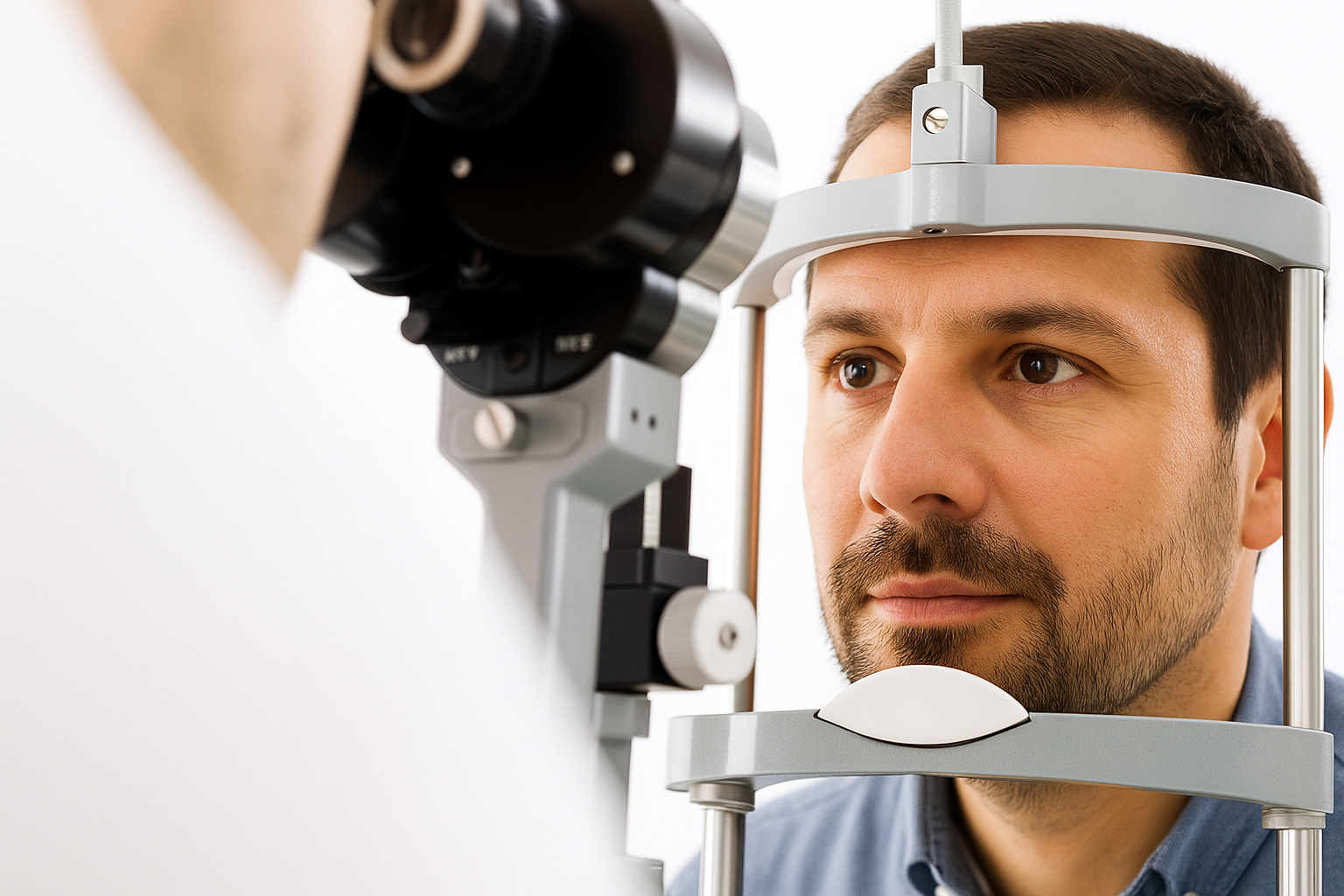
A slit-lamp microscope magnifies and illuminates the front of your eye with an intense line of light. Our doctor uses it to examine the eyelids, eyelashes, cornea, iris, lens and the fluid-filled chamber between the cornea and iris.
A dye, usually fluorescein, may be used to colour the tear film. This helps reveal damaged cells at the front of the eye. Your tears wash the dye away from the eye’s surface quite quickly.
Sometimes called ophthalmoscopy or fundoscopy, this examination allows your doctor to assess the back of the eye, including the retina, optic disc and blood vessels supplying the retina. Before the examination, drops are used to dilate the pupils, preventing them from shrinking when light is shone into the eye.
After applying the drops, the ophthalmologist uses one or more of the following techniques to examine the back of the eye.
Direct examination: The ophthalmologist uses an ophthalmoscope to shine a beam of light through the pupil and view the back of the eye. Dilating drops are not always necessary before this examination.
Indirect examination: You may sit or recline in the examination chair. The ophthalmologist examines the inside of the eye using a condensing lens and a bright light mounted on their forehead. This provides a highly detailed, three-dimensional view of the retina and other internal structures.
Non-contact tonometry measures the pressure of the fluid inside the eye, or intraocular pressure. This helps your ophthalmologist detect glaucoma, a disease that damages the optic nerve.
Several methods are available to measure intraocular pressure, including:
Non-contact tonometry: This uses a puff of air to estimate eye pressure. No instrument touches the eye, so no anaesthetic is needed. You will feel a brief pulse of air, which may be startling.
If your eye pressure is above average or the optic nerve looks unusual, your doctor may use a pachymeter, which employs sound waves to measure corneal thickness. The most common method involves applying anaesthetic drops and placing a small probe against the front surface of the eye. Measurement takes seconds.
You may need more specialised tests depending on your age, medical history and risk of developing eye disease.
Determine whether you need glasses or contact lenses.
If necessary, excimer laser surgery suitability can be assessed.
Provide comprehensive information about your eye health.
Cataracts, glaucoma, macular degeneration or diabetic retinopathy and similar conditions can be detected.
If abnormal findings are identified, further tests are planned and your doctor guides treatment.
If the examination reveals other abnormal results, our doctor may request additional tests. After these tests, the next steps in treating the condition will be discussed with you.
Dr. Hüseyin CENGİZ
Dr. Efsane H. CENGİZ
Dr. Ümit ATİK
Dr. Ali ÖZER
Dr. İdil GÖKSEL
Dr. Fatih BALTU
Dr. Ümit YAPICI
Click the useful links below: Who are our ophthalmologists? About LASIK Eye Surgery. About DCR Treatments for Blocked Tear Ducts. About Dry Eye Treatments. About Pterygium. About Glaucoma and Its Treatments. About Cataract Surgery.
Detaylı Bilgi İçin Uzmanlarımızla Görüşün
Turgut Reis Mah. 4119 Sokak No 45/A (Çetinkaya AVM Karşısı) Akdeniz/MERSİN
.
© 2026 Özel Vizyon Göz Hastanesi
Got more questions?
Learn more from one of our experts
Turgut Reis Mah. 4119 Sokak No 45/A Akdeniz/MERSİN
e-mail: info@vizyongozhastanesi.com
© 2026 Vizyon Eye Hospital
Randevu +90 324 238 83 83